Toxic Epidermal Necrolysis Treated with Intravenous Immunoglobulin and Systemic Corticosteroids: A Case Report with Favorable Clinical Response
Mariela Sanchez Lizarraga, Resident*; Rene David Sanchez Raygoza, MD; Francisco Rivera Millan, Resident; Honey Paola Meneses Cervantes, MD; Edgar Saldaña Rocha, MD; Andrea Guadalupe Zambrano Leon, MD
August 5, 2026
Abstract
Background. Stevens–Johnson syndrome (SJS) and toxic epidermal necrolysis (TEN) are severe, potentially life-threatening cutaneous adverse reactions characterized by extensive epidermal necrosis and mucosal involvement. Although treatment is primarily based on supportive care, the clinical benefit of intravenous immunoglobulin (IVIG) and systemic corticosteroids remains controversial. (1)
Methods. The objetive of this case report is to describe the therapeutic response to
combined treatment with intravenous immunoglobulin and systemic corticosteroids in a patient with toxic epidermal necrolysis.
The low incidence of Stevens–Johnson
syndrome/toxic epidermal necrolysis (SJS/TEN), its occurrence in a young patient
without underlying comorbidities, and the favorable clinical outcome following
multidisciplinary management confer clinical relevance to this case, which contributes to the available evidence on the management of these uncommon conditions.
Results. Early administration of systemic corticosteroids and intravenous
immunoglobulin, together with prompt withdrawal of the offending drug and
comprehensive supportive care, was associated with a favorable clinical course.
However, the available evidence supporting these therapies remains limited and is
derived primarily from observational studies and case reports. (3)
Conclusions. This case highlights the importance of early recognition, immediate
discontinuation of the causative agent, and multidisciplinary management in patients with SJS/TEN. It also provides additional clinical evidence supporting a therapeutic strategy that resulted in a favorable outcome, although higher-quality studies are needed to establish its efficacy. Furthermore, this case underscores the challenges of identifying the causative medication in patients receiving multiple drugs and emphasizes the importance of obtaining a thorough medication history to facilitate timely diagnosis and appropriate management.
Keywords
Toxic Epidermal NecrolysisStevens-Johnson SyndromeSevere Cutaneous Adverse ReactionsEpidermal NecrosisDrug-Related Side Effects and Adverse Reactions.
INTRODUCTION
Stevens–Johnson syndrome (SJS) and toxic epidermal necrolysis (TEN) represent a spectrum of severe, potentially life-threatening cutaneous adverse reactions characterized by massive keratinocyte apoptosis, extensive epidermal necrosis, skin detachment, and involvement of multiple mucosal surfaces and internal organs. In most cases, these conditions are triggered by medications, although they may also be associated with infections and, less frequently, with other underlying disorders. Despite their low incidence, SJS and TEN are associated with substantial morbidity and mortality, with reported mortality rates ranging from approximately 10% in SJS to more than 30% in TEN, depending on the extent of skin involvement and the patient's comorbidities. Management remains controversial, particularly regarding the use of immunomodulatory therapies such as systemic corticosteroids and intravenous immunoglobulin (IVIG), owing to the limited availability of high-quality evidence and the inconsistent findings reported across published studies. In this context, well-documented case reports continue to play an important role in expanding current knowledge of the clinical course, therapeutic strategies, and outcomes of these rare diseases. (1-2)
METHODS
The objetive of this case report is to describe the therapeutic response to combined treatment with intravenous immunoglobulin and systemic corticosteroids in a patient with toxic epidermal necrolysis.
The low incidence of Stevens–Johnson syndrome/toxic epidermal necrolysis (SJS/TEN), its occurrence in a young patient without underlying comorbidities, and the favorable clinical outcome following multidisciplinary management confer clinical relevance to this case, which contributes to the available evidence on the management of these uncommon conditions.
RESULTS
A 24-year-old previously healthy female student with no significant medical history presented to the emergency department with a 1-week history of progressive mucocutaneous lesions. Seven days prior to admission, she had been diagnosed with herpes zoster and was treated as an outpatient with acetaminophen, carbamazepine, and acyclovir.
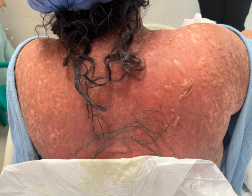
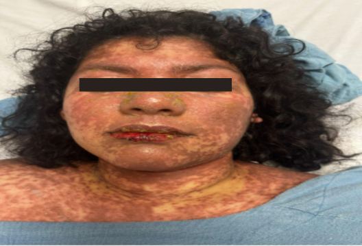
Physical examination revealed extensive, confluent erythematous maculopapular and exudative lesions involving approximately 60% of the total body surface area, associated with severe pain, pruritus, and extensive involvement of the oral, ocular, and genital mucosae. Epidermal detachment with a positive Nikolsky sign and areas of epidermal necrosis were observed, findings consistent with Stevens–Johnson syndrome/toxic epidermal necrolysis (SJS/TEN) (Figure 1 and 2).
At admission, the patient remained hemodynamically stable, without evidence of shock or acute organ dysfunction. She was promptly hospitalized under reverse isolation and placed under close clinical monitoring due to the high risk of complications associated with extensive epidermal loss.
A multidisciplinary management approach was initiated, primarily focused on intensive supportive care, including strict fluid and electrolyte management, pain control, nutritional support, temperature regulation, prevention of secondary infections, and meticulous wound care. Immunomodulatory therapy was started with high-dose intravenous methylprednisolone (100 mg/day), followed by a gradual tapering regimen over four weeks, in combination with intravenous immunoglobulin (IVIG).
During hospitalization, broad-spectrum antibiotic therapy was administered due to a documented urinary tract infection based on urinalysis findings. Additionally, topical mupirocin was prescribed for secondary impetigo. The patient also received specialized ophthalmologic evaluation and targeted management of ocular involvement, as well as continued care of mucosal and cutaneous lesions.
The clinical course was favorable, with progressive improvement characterized by stabilization of vital signs, decreased skin exudation, regression of epidermal detachment, improvement of mucosal involvement, and restoration of oral intake tolerance. Given the favorable response to treatment and the need for continued specialized multidisciplinary care, the patient was transferred to a tertiary referral center for further management and follow-up.
DISCUSSION
This case illustrates a severe presentation of a mucocutaneous syndrome in a young patient without underlying comorbidities, highlighting the unpredictable nature of these conditions. The early use of systemic corticosteroids and intravenous immunoglobulin (IVIG) remains controversial; however, in this case, their administration was associated with a favorable clinical outcome.
Several observational studies and case reports have suggested that IVIG may exert immunomodulatory effects by inhibiting Fas–Fas ligand (Fas-FasL)-mediated keratinocyte apoptosis, a pathway involved in the pathogenesis of SJS/TEN. Likewise, systemic corticosteroids may attenuate the inflammatory response when administered early and under careful monitoring. Nevertheless, the lack of high-quality randomized clinical trials and the heterogeneity of available evidence continue to limit definitive conclusions regarding the optimal immunomodulatory strategy in these patients. (3-5)
The absence of robust randomized clinical trials reinforces the value of well-documented case reports, particularly in complex and uncommon clinical scenarios. Stevens–Johnson syndrome (SJS) and toxic epidermal necrolysis (TEN) are rare, severe, and potentially life-threatening cutaneous adverse reactions that may progress rapidly, even in young patients without underlying comorbidities. (1-2) In the present case, the onset of clinical manifestations one week after the initiation of carbamazepine is consistent with the latency period described for this medication, which is recognized as one of the main triggers of SJS/TEN.
The management of SJS/TEN is primarily based on the immediate discontinuation of the causative drug, intensive supportive care, and a multidisciplinary approach. However, the role of immunomodulatory therapies remains controversial. Although intravenous immunoglobulin (IVIG) has been proposed to modulate mechanisms involved in keratinocyte apoptosis, and systemic corticosteroids may attenuate the inflammatory response when administered early in the disease course, the available evidence remains heterogeneous and is derived mainly from observational studies. Therefore, current guidelines do not provide definitive recommendations regarding their routine use. (3-6)
In the present case, early administration of intravenous methylprednisolone followed by intravenous human immunoglobulin (IVIG), combined with intensive supportive care and management of associated complications, was associated with a favorable clinical outcome. However, this outcome cannot be attributed exclusively to immunomodulatory therapy, as it likely reflects the combined effect of timely recognition, prompt withdrawal of the offending agent, and comprehensive multidisciplinary management.
Given the low incidence of SJS/TEN and the limited availability of randomized clinical trials, case reports continue to provide valuable insights into the clinical course and therapeutic approaches used in these patients, contributing to the expansion of available evidence and informing clinical practice.
CONCLUSION
Stevens–Johnson syndrome (SJS) and toxic epidermal necrolysis (TEN) are dermatologic emergencies associated with significant morbidity and mortality that require early diagnosis, immediate discontinuation of the causative drug, and a multidisciplinary approach based on intensive supportive care. In the present case, the early implementation of comprehensive management, including intravenous methylprednisolone and intravenous immunoglobulin (IVIG), was associated with a favorable clinical outcome; however, a causal relationship cannot be established due to the observational nature of this case report. This case highlights the importance of prompt recognition of these conditions and provides clinical insight into the therapeutic strategies used in a rare disease for which the optimal management approach remains under investigation.
article.figures
Figure 1. Extensive cutaneous lesions involving the dorsal region, characterized by diffuse erythema, flaccid bullae, and superficial epidermal detachment.Figure 2. Extensive mucocutaneous lesions in a patient with Stevens–Johnson syndrome/toxic epidermal necrolysis (SJS/TEN), showing diffuse facial involvement, lip erosions, and a honey-colored crusted lesion on the nasal dorsum consistent with secondary impetigo.
References
Heuer R, Paulmann M, Annecke T, Behr B, Boch K, Boos AM, Brockow K, French LE, Gille J, Gundlach V, Hartmann B, Höger P, Hofmann SC, Klein T, Lehnhardt M, Liß Y, Maier P, Mandel P, Marathovouniotis N, Marlok F, Mittelviefhaus H, Pleyer U, Pradeau M, Rall K, Rieg S, Rittner H, Sander F, Schnitzler S, Schut C, Stolle A, Vorobyev A, Wedi B, Weiss J, Zepp M, Ziemer M, Mockenhaupt M, Nast A. S3 guideline: Diagnosis and treatment of epidermal necrolysis (Stevens-Johnson syndrome and toxic epidermal necrolysis) - Part 1: Diagnosis, initial management, and immunomodulating systemic therapy. J Dtsch Dermatol Ges. 2024 Oct;22(10):1448-1466. doi: 10.1111/ddg.15515. Epub 2024 Sep 23. PMID: 39314017.
Zhou J, Wang C-P, Li J, Zhang H-L and He C-X (2024) Stevens-Johnson syndrome and toxic epidermal necrolysis associated with immune checkpoint inhibitors: a systematic review. Front. Immunol. 15:1414136. doi: 10.3389/fimmu.2024.1414136
AlFada, M., Alotaibi, H., Alsharif, S., Alani, A. H., Andrade-Miranda, A., Montesinos Guevara, C., … Amer, Y. S. (2025). Systematic review, methodological appraisal, and recommendation mapping of clinical practice guidelines for managing patients with Stevens-Johnson syndrome and toxic epidermal necrolysis. Journal of Dermatological Treatment, 36(1). https://doi.org/10.1080/09546634.2025.2467751
Frantz R, Huang S, Are A, Motaparthi K. Stevens-Johnson Syndrome and Toxic Epidermal Necrolysis: A Review of Diagnosis and Management. Medicina (Kaunas). 2021 Aug 28;57(9):895. doi: 10.3390/medicina57090895. PMID: 34577817; PMCID: PMC8472007.
Hasegawa A, Abe R. Stevens-Johnson syndrome and toxic epidermal necrolysis: Updates in pathophysiology and management. Chin Med J (Engl). 2024 Oct 5;137(19):2294-2307. doi: 10.1097/CM9.0000000000003250. Epub 2024 Sep 5. PMID: 39238098; PMCID: PMC11441865.
Chang HC, Wang TJ, Lin MH, Chen TJ. A Review of the Systemic Treatment of Stevens-Johnson Syndrome and Toxic Epidermal Necrolysis. Biomedicines. 2022 Aug 28;10(9):2105. doi: 10.3390/biomedicines10092105. PMID: 36140207; PMCID: PMC9495335.
How to cite this article
Mariela Sanchez L., Rene Sanchez R., Francisco Rivera M., et al. Toxic Epidermal Necrolysis Treated with Intravenous Immunoglobulin and Systemic Corticosteroids: A Case Report with Favorable Clinical Response. Clin Surg Rev. 2026.