
Case Report
Bilateral Posterior Shoulder Dislocation Following a Generalized Seizure: A Case Report
Mateo Alvarez, MD*; Ingrid Johansson, MD; Rohit Mehta, MD
Vol. 2, Issue 1 · May 17, 2026
Abstract
Background. Bilateral posterior shoulder dislocation is rare and frequently missed. Seizures are a classic cause.
Methods. A 34-year-old man presented after a generalized tonic-clonic seizure with bilateral shoulder pain and restricted external rotation.
Results. Radiographs and CT confirmed bilateral posterior dislocation with reverse Hill-Sachs lesions. Closed reduction under sedation was successful; function recovered with rehabilitation.
Conclusions. Bilateral posterior dislocation should be suspected after seizures when external rotation is blocked. Axillary or CT imaging avoids missed diagnosis.
Keywords
posterior shoulder dislocationbilateralseizurereverse Hill-Sachstrauma
INTRODUCTION
Posterior shoulder dislocations account for a small fraction of all shoulder dislocations and are notoriously under-recognized on standard anteroposterior films. Muscle forces during seizures can produce bilateral injury. Posterior shoulder dislocations are missed in a substantial proportion of cases at first presentation because the humeral head can appear deceptively normal on a single anteroposterior view. Bilateral injuries are almost pathognomonic of a violent, symmetric muscle contraction such as that produced by a seizure or electrocution.
METHODS
We describe the presentation, imaging, reduction and follow-up.
RESULTS
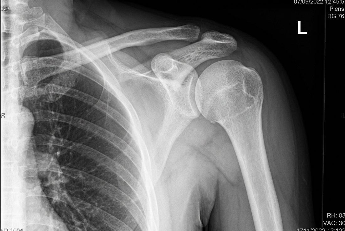
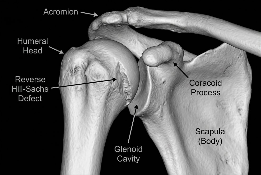
The classic 'light-bulb' sign was present on AP radiographs. CT delineated reverse Hill-Sachs defects involving less than 25% of the humeral head. Both shoulders were reduced closed and immobilized in neutral rotation, followed by physiotherapy with a good functional result at three months.
DISCUSSION
A blocked external rotation after a seizure should raise immediate suspicion. Axillary or scapular-Y views and CT prevent the delayed diagnosis that predisposes to recurrent instability and arthropathy. Prompt recognition depends on clinical suspicion and adequate imaging, including axillary or scapular-Y views and, when needed, computed tomography to quantify the reverse Hill-Sachs defect. Early closed reduction and immobilization in neutral or slight external rotation optimize the chance of stable, functional recovery.
CONCLUSION
Awareness and appropriate imaging enable prompt reduction and good outcomes in bilateral posterior shoulder dislocation.
article.figures
References
- Cicak N. Posterior dislocation of the shoulder. J Bone Joint Surg Br. 2004;86(3):324-332.
- Robinson CM, Aderinto J. Posterior shoulder dislocations and fracture-dislocations. J Bone Joint Surg Am. 2005;87(3):639-650.
- Brackstone M, Patterson SD, Kertesz A. Triple 'E' syndrome: bilateral posterior fracture-dislocation. Can J Neurol Sci. 2001;28(2):163-165.