Late subacute peripancreatic hematoma secondary to walled-off necrotizing pancreatitis: a case report
Enrique Acuña Ortega, Resident*; Cinthya Viridiana Hernández Muñoz, MD; Vanessa Osuna Velarde, MD; Jonatan Ezequiel Camacho Lopez, MD; Emiliano López León, MD; Pablo Maggiani Aguilera, MD; Andrea Guadalupe Zambrano León, MD; Brandon Javier Uriarte Zamora, MD; Edgar Saldaña Rocha, MD; Jose Alejandro Gomez, MD; Blanca Estela Melendez Acosta, MD
August 24, 2026
Abstract
A 38-year-old patient with a history of biliary acute pancreatitis treated with cholecystectomy developed epigastric pain and anemia two months after the surgical intervention. Imaging studies revealed peripancreatic hemorrhagic collections, compatible with intra-abdominal hemorrhage. Due to clinical deterioration, an exploratory laparotomy was performed, revealing approximately 2000 mL of hemoperitoneum without an active bleeding source. Analysis of the drainage fluid showed elevated amylase and lipase levels, confirming the presence of an internal pancreatic fistula. This case illustrates a rare late hemorrhagic complication of pancreatitis and highlights the importance of clinical suspicion and imaging for timely diagnosis.
Delayed intra-abdominal hemorrhage is a rare clinical entity, most commonly associated with abdominal trauma, anticoagulation therapy, or underlying coagulation disorders. In exceptional cases, it has been reported as a complication of inflammatory abdominal conditions, including acute pancreatitis, particularly in necrotizing pancreatitis or in pancreatic pseudocysts complicated by vascular erosion. Nevertheless, the occurrence of a delayed hemorrhagic collection secondary to focal necrotizing inflammatory pancreatitis, without a history of trauma or hemorrhagic risk factors, represents an exceedingly rare clinical presentation. (1) Hemorrhagic complications associated with acute pancreatitis are uncommon but potentially life-threatening and are generally related to vascular erosion secondary to the proteolytic activity of pancreatic enzymes. Atypical hemorrhagic manifestations have been described in the literature, including subcapsular splenic or hepatic hematomas, as well as intrahepatic hemorrhage, which may develop in the setting of acute or necrotizing pancreatitis. (2,3) However, these presentations are exceedingly rare, and their diagnosis often represents a clinical challenge due to their low incidence and the nonspecific nature of the initial findings. In particular, the formation of intra-abdominal hemorrhagic collections distant from the pancreatic parenchyma has been scarcely reported. In this context, the aim of the present report is to describe a case of a hemorrhagic collection secondary to walled-off necrotizing pancreatitis with the development of an internal pancreatic fistula, contributing to the recognition of this uncommon complication and highlighting the importance of early diagnostic suspicion.
METHODS
A clinical case report was conducted to document a rare late hemorrhagic complication associated with walled-off necrotizing pancreatitis, characterized by the development of a peripancreatic hematoma and a concomitant internal pancreatic fistula. This case was considered clinically relevant because of the uncommon nature of this presentation, its nonspecific clinical manifestations, and the diagnostic challenge it may represent, particularly in the absence of a history of trauma, anticoagulant use, or other recognized hemorrhagic risk factors. A retrospective review of the patient’s medical record was performed, including relevant medical history, clinical course, laboratory findings, imaging studies, intraoperative findings, biochemical analysis of the drainage fluid, treatment, and subsequent clinical follow-up. Abdominal magnetic resonance imaging and contrast-enhanced computed tomography findings were reviewed, together with the findings obtained during exploratory laparotomy, to characterize the hemorrhagic collection and its relationship with the underlying pancreatic inflammatory process. The biochemical characteristics of the postoperative drainage fluid were also evaluated to support the diagnosis of an internal pancreatic fistula. The case was documented for academic and scientific purposes, with the aim of increasing awareness of this uncommon complication and emphasizing the importance of maintaining a high index of clinical suspicion and timely use of appropriate imaging modalities in patients with atypical or delayed complications of necrotizing pancreatitis.
RESULTS
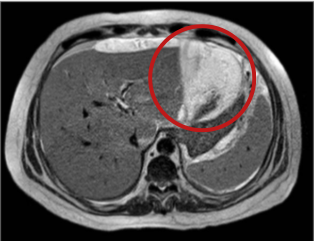
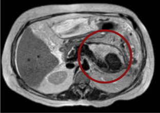
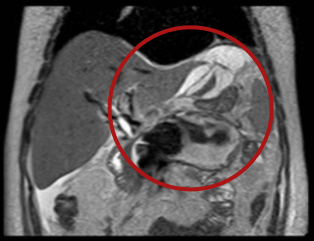
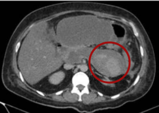
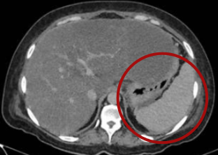
A 38-year-old female patient was referred to a secondary level hospital for evaluation of acute abdominal syndrome. Her relevant medical history included a previous hospitalization for mild acute pancreatitis, classified according to the revised Atlanta criteria, of biliary etiology secondary to biliary tract obstruction. During that episode, early cholecystectomy was performed in accordance with the recommendations of the American Gastroenterological Association guidelines. Intraoperatively, the gallbladder was classified as Parkland grade II, with no surgical complications. The patient had an uneventful postoperative course and was discharged without evidence of complications. Approximately 60 days after the initial hospitalization, the patient presented with a new episode characterized by epigastric pain, accompanied by nausea and subsequent vomiting on five occasions. Her condition progressively deteriorated over the following hours, with increasing abdominal pain. Upon admission, physical examination revealed tenderness to superficial palpation and a palpable mass in the epigastric region and right hypochondrium, without muscular rigidity or evidence of periumbilical or flank ecchymosis. Initial laboratory studies showed a hemoglobin concentration of 9.2 g/dL, serum glucose of 230 mg/dL, hyperbilirubinemia with total bilirubin of 1.44 mg/dL, direct bilirubin of 0.65 mg/dL, and indirect bilirubin of 0.80 mg/dL, as well as elevated alkaline phosphatase levels of 1,657 IU/L. C-reactive protein (CRP) was elevated at 12.35 mg/L. Serum amylase and lipase levels were requested, revealing elevated concentrations of lipase (1,345 IU/L) and amylase (845 IU/L). Forty-eight hours after admission, the patient showed clinical deterioration, with a marked increase in CRP levels to 161 mg/dL and a decrease in hemoglobin concentration to 7.2 g/dL, requiring transfusion of three units of packed red blood cells. Given the laboratory findings and clinical progression, a non-contrast magnetic resonance cholangiopancreatography (MRCP) was performed, revealing the following findings: a septated collection with hemosiderin remnants in the gastrohepatic space measuring 15 × 15 × 6 cm (approximately 700 mL) (Figure 1), and a retroperitoneal collection adjacent to the pancreatic tail measuring 9 × 6 × 7 cm (approximately 240 mL) (Figure 2), both findings compatible with organized hematomas. Additional findings included perihepatic free fluid, right basal atelectasis, left pleural effusion, and an interloop collection in the left flank measuring 6 × 5 cm (Figure 3). To assess pancreatic viability and further characterize the collections, a contrast-enhanced abdominal computed tomography (CT) scan was obtained. The study demonstrated findings consistent with focal pancreatitis, including absence of contrast enhancement of the pancreatic parenchyma during both arterial and venous phases (Figure 4). An encapsulated heterogeneous collection adjacent to the pancreas with hemorrhagic and inflammatory content was identified (Figure 5), suggestive of a hematoma with an estimated chronicity of 2–7 days, with possible communication toward the perihepatic Morrison’s pouch and the lesser sac. During hospitalization, the patient developed intermittent fever episodes with peaks of up to 38.5 °C. Due to their temporal association with blood product administration, these episodes were attributed to a probable non-hemolytic febrile transfusion reaction. The case was evaluated by the Hematology Department, which recommended symptomatic management and prophylactic measures in case additional transfusions were required. The patient showed adequate clinical response, with resolution of fever within the following 48 hours. On the eighth day of hospitalization, after hemodynamic stabilization and optimization of hemoglobin levels, the patient was taken to the operating room for exploratory laparotomy. During the procedure, approximately 2,000 mL of chronic-appearing hemorrhagic fluid undergoing coagulation was identified, predominantly located in the suprahepatic region (left hepatic lobe), anterior to the stomach, within interloop spaces, and in the lesser sac. Additionally, approximately 100 mL of inflammatory fluid was observed. No active bleeding source was identified during intraoperative exploration. Extensive surgical lavage of the abdominal cavity was performed, and Penrose drains were placed for postoperative monitoring and control. Subsequently, the patient remained under close postoperative surveillance. Biochemical analysis of the fluid obtained through the Penrose drain revealed elevated amylase (8,788 U/L) and lipase (>25,000 U/L) concentrations, findings consistent with a pancreatic leak. The patient's clinical course was favorable, with progressive improvement, adequate oral intake tolerance, hemodynamic stability, and no evidence of infection. These findings allowed hospital discharge with outpatient follow-up for monitoring of drain output and clinical surveillance. At the three-week follow-up evaluation, the patient was asymptomatic and showed no evidence of complications; therefore, she was discharged from medical care with a diagnosis of a type A pancreatic fistula.
DISCUSSION
Acute pancreatitis is one of the most common causes of hospitalization due to gastrointestinal disease and may progress to multiple local and systemic complications. Among these, vascular complications represent an uncommon but potentially fatal group, primarily resulting from enzymatic autodigestion of pancreatic tissue and adjacent vascular structures. This inflammatory process may lead to weakening of the vascular wall, pseudoaneurysm formation, or vessel rupture, resulting in intra-abdominal hemorrhage. Vascular complications have been estimated to occur in approximately 1–23% of patients with pancreatitis, whereas hemorrhagic arterial complications occur in approximately 1.3–10% of cases. (4-9) Furthermore, when significant bleeding occurs, mortality rates may reach 34–52%, emphasizing the importance of early recognition and timely management. (10)
In most reported cases, intra-abdominal hemorrhage is associated with abdominal trauma or postoperative complications. (11) Consequently, during the initial evaluation of the present case, a complication related to the previous surgical procedure was considered the primary diagnostic hypothesis. However, this possibility was definitively excluded based on the intraoperative findings. The differential diagnosis included mesenteric vascular disorders, anticoagulant therapy, and neoplastic processes. Nevertheless, the absence of relevant medical history, together with the lack of compatible clinical, biochemical, and radiological findings, allowed these etiologies to be ruled out. The nonspecific clinical presentation, combined with the limited diagnostic sensitivity of medical history and physical examination in this setting, highlights the need for advanced imaging modalities, including computed tomography and magnetic resonance imaging, for the timely detection of hemorrhagic complications associated with pancreatitis. (7-8, 12-13)
During the postoperative period, biochemical analysis of the fluid obtained through the Penrose drain revealed markedly elevated amylase and lipase concentrations, directing the pathophysiological assessment toward the presence of a pancreatic fistula. According to the International Study Group on Pancreatic Fistula (ISGPF) classification, the condition was initially considered a type C pancreatic fistula due to its significant clinical impact, characterized by the development of an extensive intra-abdominal collection, the requirement for surgical intervention, and its influence on the patient's clinical course. These findings suggest focal erosive damage of the peripancreatic vascular structures secondary to walled-off necrotizing pancreatitis localized in the pancreatic head, with extravasation of pancreatic secretions into the peritoneal cavity. Anatomically, the vessels most frequently involved include the celiac trunk, superior mesenteric artery, and, predominantly, the splenic artery and its distal branches. Subsequently, following favorable clinical evolution and progressive resolution of the condition, the fistula was reclassified as a less severe type A pancreatic fistula during follow-up. (14-15)
Additionally, intracavitary hemorrhage may occur when vascular rupture extends into a pancreatic pseudocyst or an encapsulated necrotic collection, appearing on imaging studies as hyperattenuating or hyperintense areas depending on the modality used. (4, 6, 9, 12-15) In the present case, non-contrast magnetic resonance cholangiopancreatography demonstrated a peripancreatic collection with a thin, well-defined wall, heterogeneous peripancreatic tissue, and alterations in the morphology of the pancreatic tail. These radiological findings, together with the clinical evolution, supported the diagnosis of a focal and localized vascular erosive injury.(12, 13, 16) Furthermore, Thomas R. Gadacz et al. performed a meta-analysis on vascular lacerations associated with pancreatitis, reporting an incidence of hemorrhagic complications ranging from 1.7% to 2.5%. (17, 18) In a review of 44 cases, the splenic artery and gastroduodenal artery were identified as the most frequently affected vessels, with incidences of 34% and 9%, respectively. In 28% of cases, the injured vessel could not be identified, which is consistent with the present case, in which no vascular injury was identified during surgical exploration. Notably, these complications were more frequently observed in patients with chronic pancreatitis of alcoholic etiology, accounting for up to 75% of reported cases. (19) In 2020, Vikas Gupta et al. published a meta-analysis focused on hemorrhagic complications associated with acute pancreatitis. Among 183 cases of severe acute pancreatitis analyzed, only 13% (24 patients) developed hemorrhagic complications. Of these, 50% presented with intra-abdominal or intraluminal hemorrhage, 52% experienced massive bleeding, and the remaining patients developed mild hemorrhage. Sixteen cases were classified as de novo hemorrhage, whereas eight were categorized as postoperative hemorrhage. Based on these findings, the authors proposed a clinical classification according to bleeding location and severity: intraluminal hemorrhage (presenting as hematemesis or melena), intra-abdominal hemorrhage (identified as a hemorrhagic collection on imaging studies), massive hemorrhage (defined as a hemoglobin decrease >2 g/dL or hemodynamic instability), and mild hemorrhage. According to this classification, the present case was categorized as a de novo massive hemorrhage, as the patient had not undergone a previous surgical procedure directed toward the management of peripancreatic hemorrhage. Notably, within that study, only four cases presented with these characteristics; two were associated with pseudoaneurysm formation, whereas the underlying etiology could not be determined in the remaining two cases. (20)
CONCLUSION
The management of this case represented a considerable diagnostic and therapeutic challenge, mainly due to the atypical location of the hematoma, limitations in access to advanced imaging modalities such as angiography, and the prolonged clinical course. Ultimately, it was concluded that the late subacute hemorrhagic event was secondary to walled-off necrotizing pancreatitis localized in the pancreatic head, documented by magnetic resonance imaging, which resulted in peripancreatic vascular injury and the concomitant development of a type C internal pancreatic fistula as a complication of a localized inflammatory process in this region. (6, 14-15) Spontaneous peripancreatic hemorrhagic collections associated with acute pancreatitis represent an uncommon complication with a frequently nonspecific clinical presentation, which may delay the timely recognition of hemorrhagic events. Therefore, maintaining a high index of suspicion is essential in patients presenting with clinical deterioration or atypical findings during the course of the disease, even in the absence of classical criteria for severe or necrotizing acute pancreatitis. Early recognition of these complications allows optimization of diagnostic and therapeutic strategies, potentially improving patient outcomes.
article.figures
Figure 1. Non-contrast axial T2-weighted abdominal magnetic resonance imaging (MRI) showing a perihepatic collection with heterogeneous signal intensity and predominantly hyperintense characteristics.Figure 2. Non-contrast axial T2-weighted abdominal MRI demonstrating a peripancreatic collection with a thin, well-defined wall, heterogeneous peripancreatic tissue, and associated alteration in the morphology of the pancreatic tail.Figure 3. Non-contrast coronal T2-weighted abdominal MRI showing a peripancreatic collection associated with a perihepatic collection extending toward the left subdiaphragmatic space.Figure 4. Portal venous phase contrast-enhanced abdominal computed tomography (CT) demonstrating a peripancreatic collection with a well-defined wall and adjacent peripancreatic tissue showing avid contrast enhancement.Figure 5. Portal venous phase contrast-enhanced abdominal CT showing a peripancreatic collection with homogeneous hypodense content and a thin, well-defined wall, causing displacement of the lesser gastric curvature.
References
Lin CK, Chen CH, Yeh CH, Lin SL, Tsang YM, Sheu JC. Intrahepatic hemorrhage and subcapsular hematoma developing in acute pancreatitis. Hepatogastroenterology. 2003 Mar-Apr;50(50):571-3. PMID: 12749275.
Duncan DP, Briese AK, Niemiec S, White R, Picel AC, Hahn ME. Subcapsular hematoma resulting in hepatic ischemia as a complication of necrotizing pancreatitis. Radiol Case Rep. 2020 Jan 20;15(4):316-320. doi: 10.1016/j.radcr.2019.12.021. PMID: 31988681; PMCID: PMC6971341.
Fenando A, Tatineni S, Raziq FI, Alratroot A. Subcapsular haematoma of the spleen complicating acute pancreatitis. BMJ Case Rep. 2019 Sep 11;12(9):e231716. doi: 10.1136/bcr-2019-231716. PMID: 31511275; PMCID: PMC6738698.
Singh, Amandeep MD1; Aggarwal, Manik MD2; Garg, Rajat MD1; Walsh, Matthew MD3; Stevens, Tyler MD1; et al. Spontaneous Internal Pancreatic Fistulae Complicating Acute Pancreatitis. The American Journal of Gastroenterology 116(7):p 1381-1386, July 2021. | DOI: 10.14309/ajg.0000000000001282
BanksPA,FreemanML;Practice Parameters Committee of the American College of Gastroenterology. Practice guidelines in acute pancreatitis. Am J Gastroenterol. 2006;101(10):2379–400. https://doi.org/10.1111/j. 1572-0241.2006.00856.x
Evans RP, Mourad MM, Pall G, Fisher SG, Bramhall SR. Pancreatitis: preventing catastrophic haemorrhage. World J Gastroenterol. 2017;23(30):5460–8. https://doi.org/10.3748/wjg.v23.i30.5460
FlatiG, Andrén-SandbergA, LaPintaM, PorowskaB, CarboniM. Potentiallyfatal bleeding in acute pancreatitis: pathophysiology, prevention, and treatment. Pancreas. 2003;26(1):8–14. https://doi.org/10.1097/ 00006676-200301000-00002
WeiAL, Guo Q, Wang MJ, Hu WM, Zhang ZD. Early complications after interventions in patients with acute pancreatitis. World J Gastroenterol. 2016;22(9):2828–36. https://doi.org/10.3748/wjg.v22.i9.2828
ChenY, ZhouJ, LiG, TongZ, DongJ, PanY, et al. Earlyspontaneous abdominal bleeding is associated with poor outcome in moderate to severe acute pancreatitis patients: a propensity matched study. Sci Rep. 2017;7: 42607. https://doi.org/10.1038/srep42607
Sanchez Cruz C, Abera Woldehana N, Ponce-Lujan L, Shettywarangale P, Shekhawat P, da Silva N, et al. Comprehensive Review of Surgical and Radiological Management of Hemorrhagic Pancreatitis: Current Strategies and Outcomes. Cureus. 2024 Jul 21;16(7):e65064. doi: 10.7759/cureus.65064. PMID: 39171005; PMCID: PMC11336159.
KalasMA, LeonM, Chavez LO, Canalizo E, Surani S. Vascular complications of pancreatitis. World J Clin Cases. 2022;10(22):7665–73. https://doi.org/10.12998/wjcc.v10.i22.7665
Claudio Bassi, Christos Dervenis, Giovanni Butturini, Abe Fingerhut, Charles Yeo, Jakob Izbicki, et al, Postoperative pancreatic fistula: An international study group (ISGPF) definition, Surgery, Volume 138, Issue 1, 2005, Pages 8-13, ISSN 0039-6060, https://doi.org/10.1016/j.surg.2005.05.001.
Duncheskie R, Anjum F. Pancreatic Fistula. [Updated 2025 Feb 22]. In: StatPearls [Internet]. Treasure Island (FL): StatPearls Publishing; 2025 Jan-. Available from: https://www-ncbi-nlm-nih-gov.pbidi.unam.mx:2443/books/NBK560871/
DörffelT, WruckT, RückertRI, RomaniukP, DörffelQ, WermkeW. Vascularcomplicationsinacutepancreatitis assessed by color duplex ultrasonography. Pancreas. 2000;21(2):126–33. https://doi.org/10.1097/ 00006676-200008000-00004
BanksPA, Bollen TL, Dervenis C, Gooszen HG, Johnson CD, Sarr MG, et al. Classi cation of acute pancreatitis: 2012– revision of the Atlanta classi cation and de nitions by international consensus. Gut. 2013;62(1): 102–11. https://doi.org/10.1136/gutjnl-2012-302779 88
Gupta V, Krishna P, Kochhar R, Yadav TD, Bargav V, Bhalla A, et al. Hemorrhage complicating the course of severe acute pancreatitis. Ann Hepatobiliary Pancreat Surg. 2020;24(3):292–300. https://doi.org/10.14701/ ahbps.2020.24.3.292
Balachandra S, Siriwardena AK. Systematic appraisal of the management of the major vascular complications of pancreatitis. Am J Surg. 2005;190(3):489–95. https://doi.org/10.1016/j.amjsurg.2005.03.009
Balthazar EJ. Acute pancreatitis: assessment of severity with clinical and CT evaluation. Radiology. 2002; 223(3):603–13. https://doi.org/10.1148/radiol.2233010680
How to cite this article
Enrique Acuña O., Cinthya Hernández M., Vanessa Osuna V., et al. Late subacute peripancreatic hematoma secondary to walled-off necrotizing pancreatitis: a case report. Clin Surg Rev. 2026.